Evaluation of the cytotoxic effects of compounds on single spheroids

Introduction

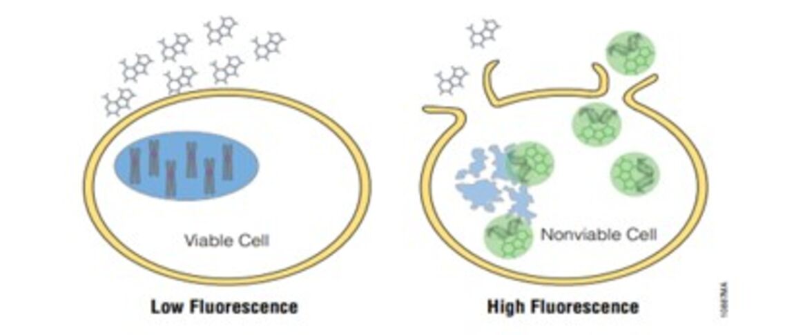
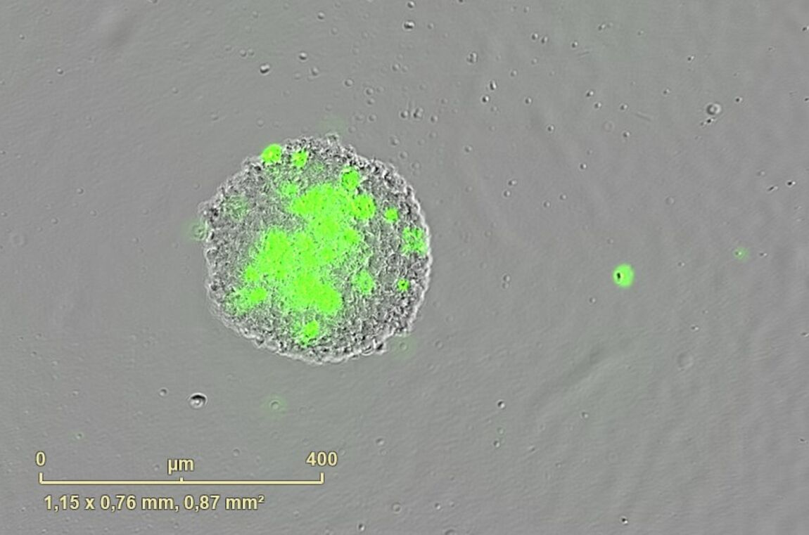
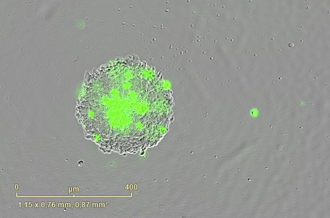
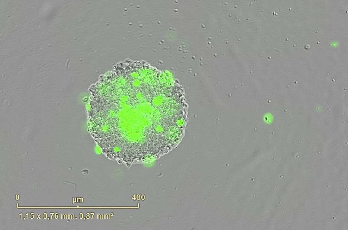
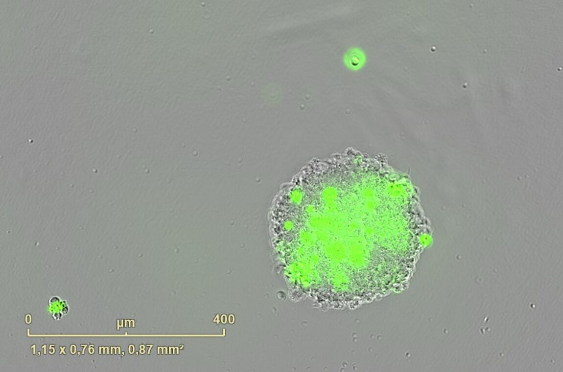
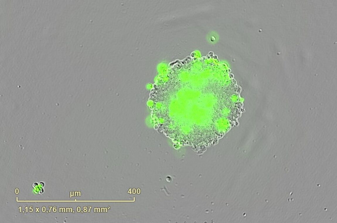
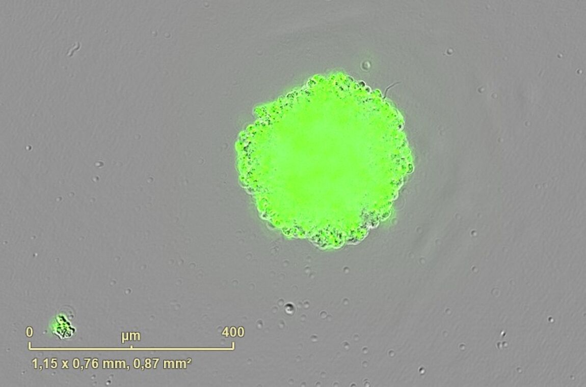
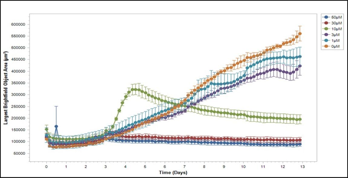
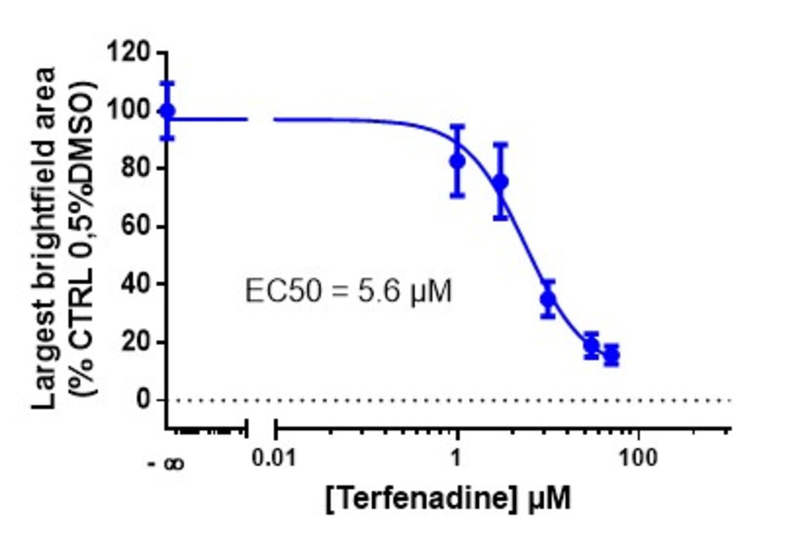
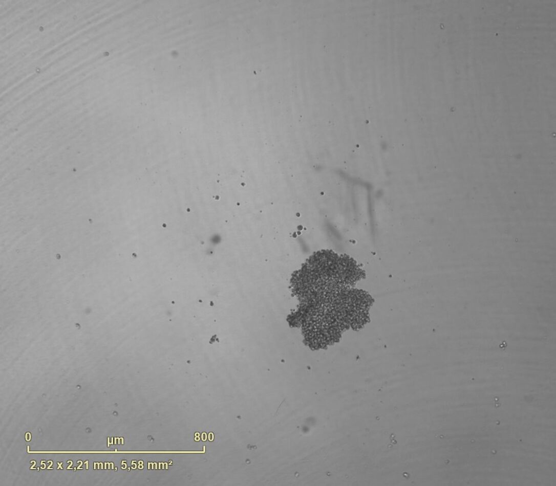
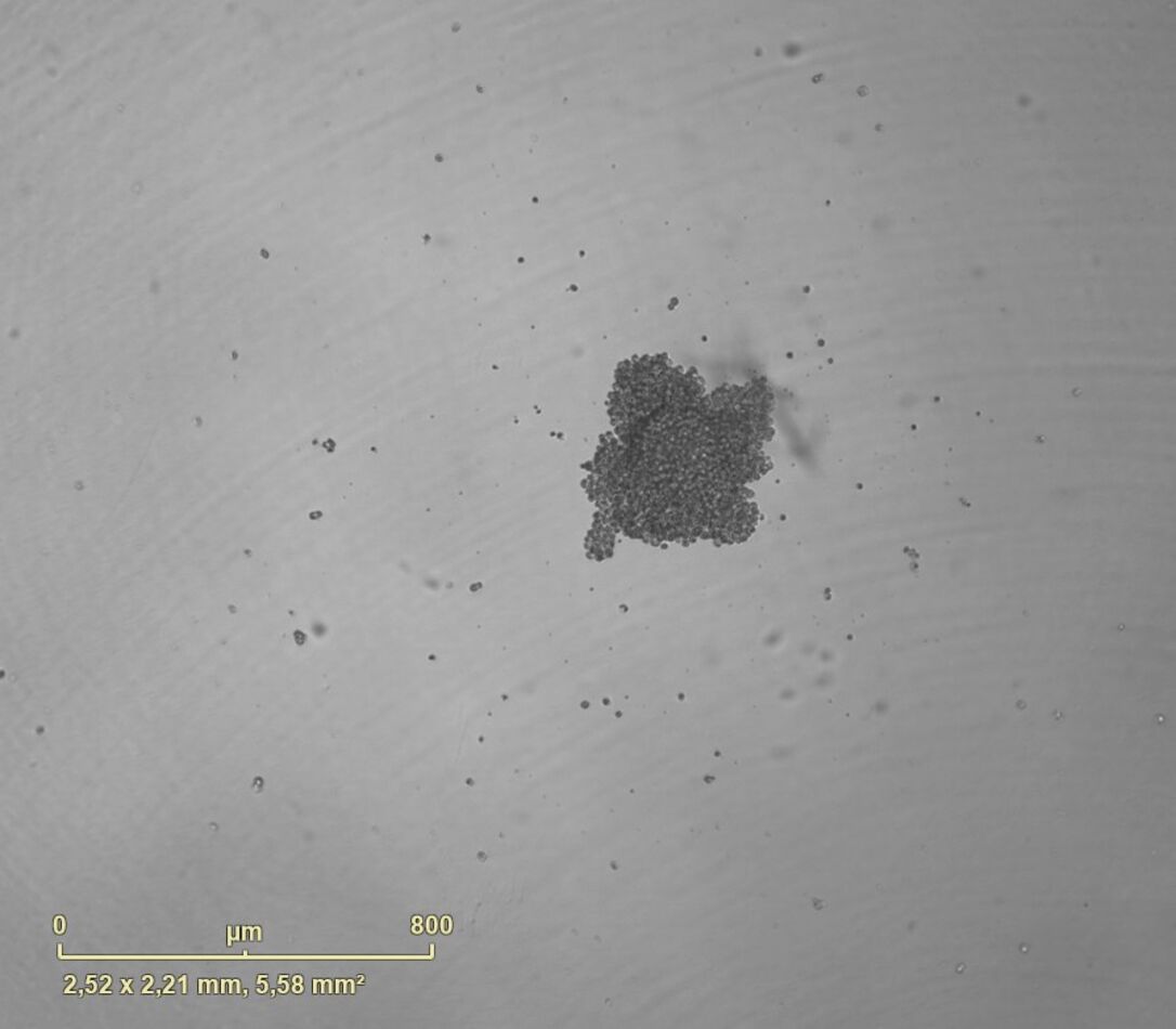
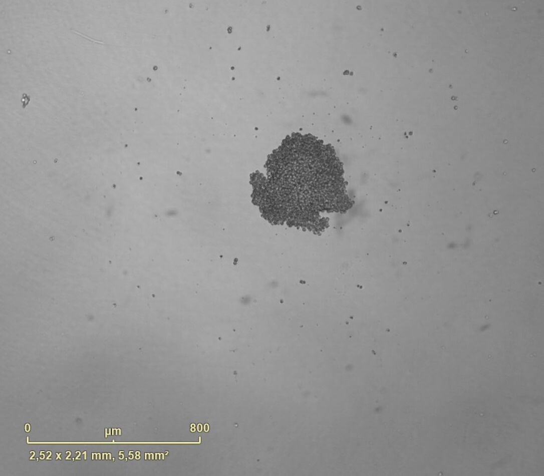
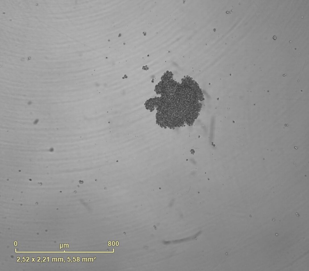
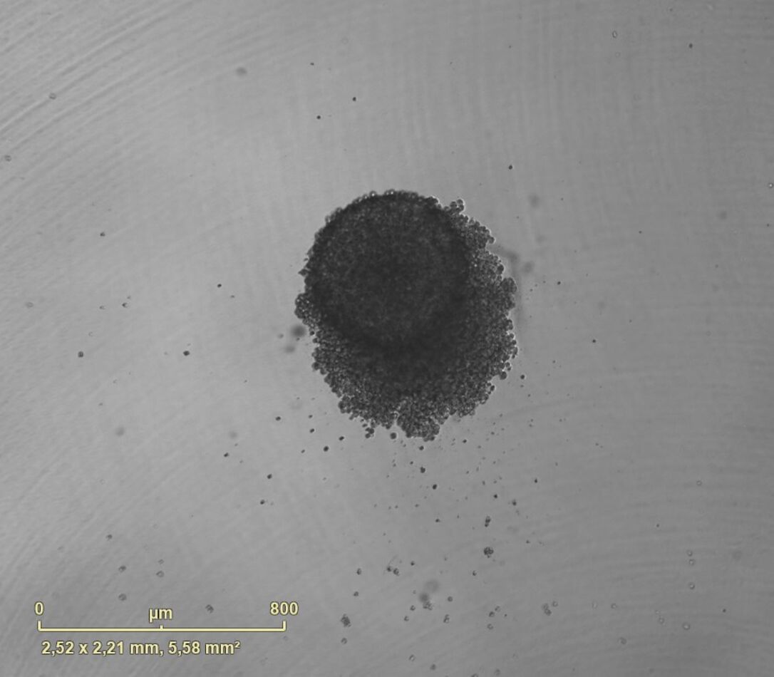
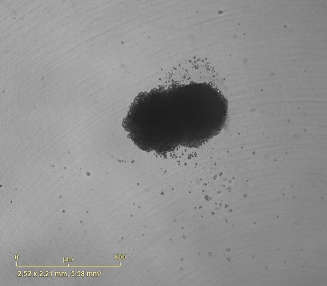
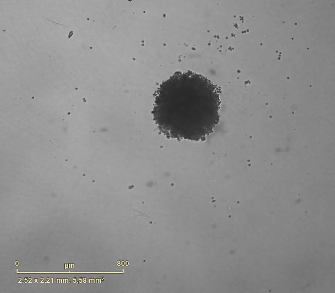
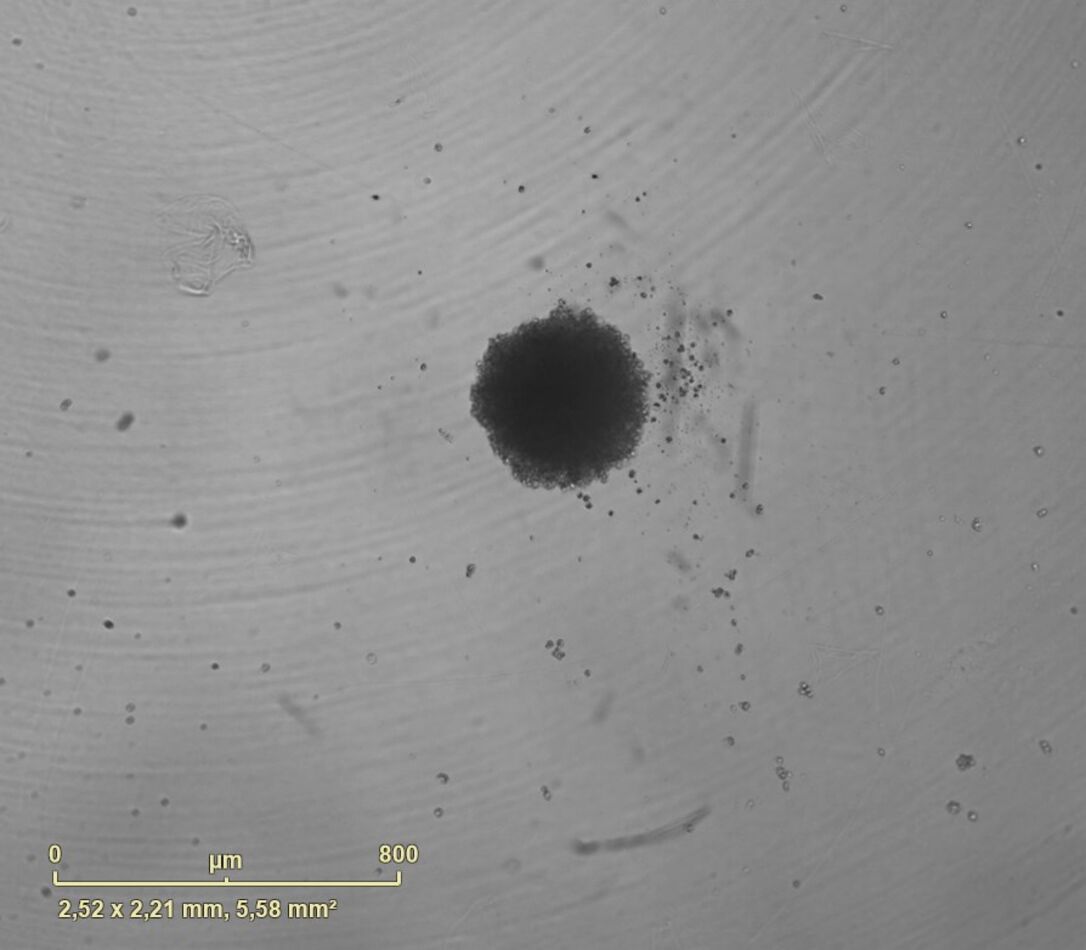
PCBIS proposes a miniaturized microplate assay on spheroids from malignant glioneuronal tumor cells for the evaluation of the cytotoxic effect of compounds. The cytotoxic effect can be quantified by monitoring the viability of spheroids or by monitoring the growth of spheroids.

Principle

1. Measurement of spheroid viability by fluorescence

The CellTox-Green assay (PROMEGA) allows to quantify the viability of spheroids over time under conventional culture conditions in a controlled atmosphere using the Incucyte S3 (Essen Bioscience /Sartorius).

The fluorescent molecule cyanine from the kit stains only DNA of non-viable cells.                     

The fluorescent signal (ex 512 nm / em 532 nm) produced by cyanine is proportional to the cell cytotoxicity.

2. Measurement of spheroid growth by phase contrast

Spheroid growth is quantified in real time using the Incucyte S3 (Essen Bioscience /Sartorius) which allows the measurement of spheroid area.

Protocol

Format96 wells  - round bottom plate
Cellular  model

Malignant glioneuronal tumors
possibility to work on other cells

Reference moleculeTerfenadine
Incubation

15 hours at 37°C under 5% CO2 (viability monitoring by fluorescence)
10 days at 37°C under 5% CO2 (growth monitoring by phase contrast)

possibility to adapt the incubation time to the biological model

Viability assayCellTox-Green (PROMEGA) kinetic and/or CellTiter-Glo (PROMEGA) 1 point
Detection

Incucyte® S3 (SARTORIUS) - phase contrast / Fluorescence and/or Luminescence measurement

Notes :

  • Allow 50 µL at 10 mM or 3 mg of sample to test 5 concentrations (3 points/concentration)

Recommendation :

  • Ensure sample solubility in a solvent compatible with cell culture


1. Monitoring of spheroid viability by fluorescence

2. Monitoring of spheroid growth by phase contrast

All models can be adapted to your request.