Intracellular calcium flux measurement

Introduction

Calcium is involved in many physiological processes (neurotransmitter release, muscle contraction, blood coagulation...) and in cell signalling where it acts as a second messenger following the activation of heterotrimeric Gq proteins via G protein coupled receptors.
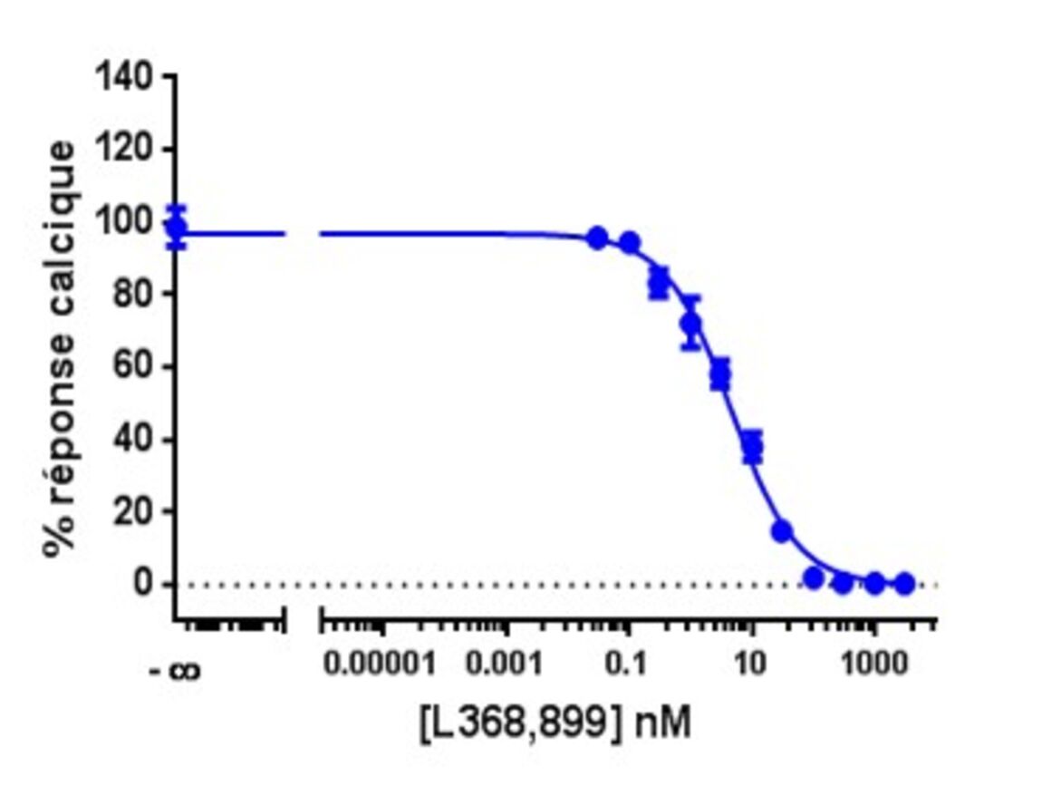
This test allows dynamic monitoring of variations in intracytoplasmic calcium concentration in living cells.

Principle

Calcium flux measurements are performed using a calcium-sensitive fluorescent probe (Indo1 or Fluo4). Depending on the nature of the probe, its binding to calcium induces a variation in its fluorescence intensity and possibly a shift in its emission spectrum :

The probes are made permeable by the addition of an acetomethylester (AM) moiety, and in this form can pass the plasma membrane of cells.  The AM part is released inside the cell by intracellular esterases.
The measurements are performed on a semi-robotic reader (Flexstation3® Molecular Devices) which allows real-time detection of intracellular calcium release.

Protocol

Format     96 or 384 well format
Cellular model     HEK293 in suspension  possibility to work on other cell lines
Calcium probe

      Indo1-AM (λex338nm / λem1401nm, λem2475nm)

      Fluo4-AM (λex494nm / λem516nm)

Loading of cells with the probe  

Cell detachment and distribution in the plate

Baseline signal measurement

Treatment of cells with compounds of interest + measurement

Up to 3 consecutive treatments possible.

Notes :

  • Quantity of cells expressing the receptor of interest (transient or stable line) to be defined during the test validation
  • Allow 2 to 3 mg of compounds to be tested

Recommendation :

  • Ensure solubility of the sample in a solvent compatible with cell culture

Stimulation of the oxytocin receptor and measurement of the associated calcium signal

Format : 384 wells
Cells : HEK293 overexpressing oxytocin receptor

All the models can be adapted to your needs.