Measurement of receptor-ligand binding by HTRF

Introduction

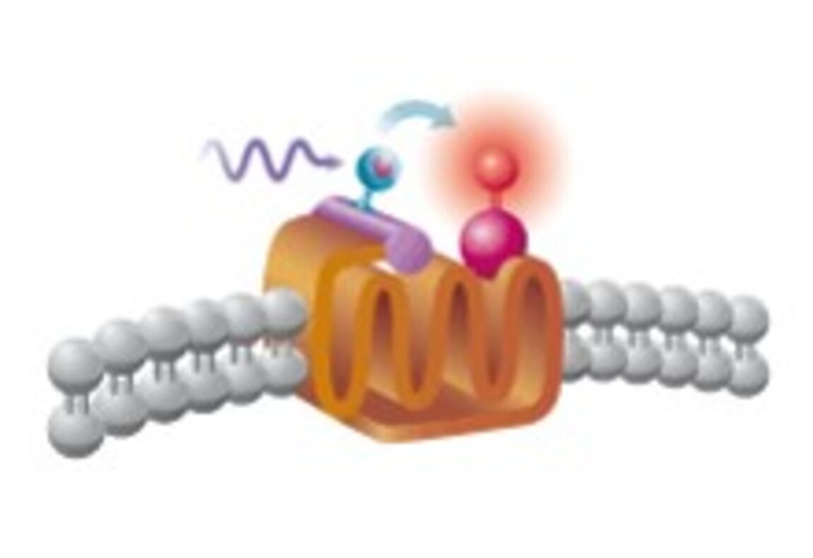
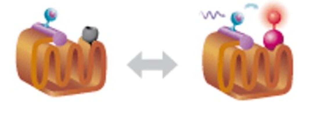
Similar to FRET, the Homogeneous Time Resolved Fluorescence (HTRF) technology allows to measure the binding between a membrane receptor fused to a fluorophore and a ligand fused to another fluorophore.
It represents a good alternative to FRET or radioactive tests. Among other benefits, it enables to overcome the transient auto-fluorescence of reagents and consumables that contribute to the background noise, by performing a measurement shifted in time by 50 to 150 microseconds after the excitation of the donor fluorophore (CISBIO).

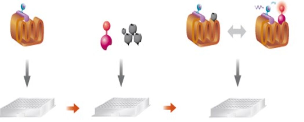
The binding is measured on living whole cells.
The assay is performed under homogeneous conditions and therefore does not require any washing.

Principle

The cells overexpress the receptor fused to a SNAP-Tag, CLIP-Tag or Halo-Tag.
These tags covalently interact with terbium or europium derivative used as donor fluorophores.
Plasmid constructs or already transfected cells are available at Cisbio International.

The detection of the binding of a ligand fused to a compatible acceptor fluorophore is performed by a microplate reader.

By competition, it is possible to detect the binding of an unlabeled molecule to the receptor.

www.cisbio.com

Protocol

To be adapted according to the test

Format384-well
Cellular modelsuspension of HEK 293  

Possibility to work on other lines

Cell labeling    Possibility to prepare a labeled frozen stock

Distribution of the cells in the plate

Addition of the fluorescent ligand and the unlabeled compound

Incubation
DetectionExcitation wavelength : 337 nm
Emission wavelength 1 : 665 nm
Donor fluorophore emission
Emission wavelength 2 : 615 nm
Acceptor fluorophore emission

Notes :

  • The quantity of cells expressing the receptor of interest to use in the experiment (transient or stable line) needs to be evaluated during assay validation.
  • Prepare 20 µL of labeled ligand in 10 mM solution in DMSO.

Measurement of V1A receptor ligand binding

Cells  : HEK293 overexpressing the V1A-SNAPTag receptor from Cisbio (reference C1PU1V1A)

Fluorescent ligand from Cisbio (reference L0003RED), 1 nM

Analysis : S = Signal at 665 nm / Signal at 615 nm x 10

All models can be adapted to your request.