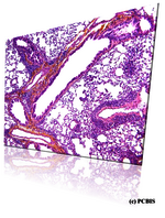
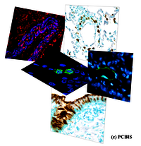
Histology and immunostaining

The study in humans and animals (mice, rat, etc.) may require histological exploration of cells or tissues. We bring you our technical and technological expertise in the various fields of histology for the development of your project.

Fixing, decalcification and paraffin impregnation

We have a Leica Pearl® automaton and standardized protocols adapted to different tissues and organs and allowing fine sections to be made with a microtome (3 to 6 µm).

Cytocentrifuge

We have a Cytospin® 4 (Shandon), a useful cytocentrifuge for the preparation of non-adherent cells on a slide that can be used later for immunolabeling (ICC).

Tissue staining

Tissue staining is performed using a Shandon Varistain XY automaton suitable for staining a large number of slides in 1 pass (60 slides) and ensures good reproducibility. A wide range of stains is offered such as: Hematoxylin Eosin Safron (HES), Trichrome de Gomori (collagen), luna (eosinophils), toluidine blue and alciane/safranin blue (Mastcells), Periodic acid schiff (Mucus).

Immunolabelling on cells and tissues

We use the Shandon Sequenza system with cover plate to develop and standardize immunohistochemistry and immunofluorescence labeling of proteins of interest.

Image analysis

We have a Leica macroscope and microscopes equipped with a high-sensitivity 12.8 Megapixel camera (Olympus, 4140 x 3096 pixels) allowing semi-automated analysis in brightfield and fluorescence.